

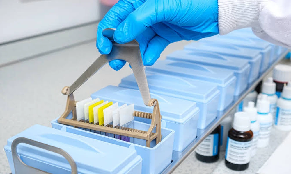
Look at almost any pathology slide and the deep blue-purple nuclei you see come from one old, dependable dye. In many labs that colour comes from Harris hematoxylin solution, a formulation that has been part of routine H&E work for more than a century. It gives crisp nuclear detail, and it rewards the staff who understand what is happening in the bottle and on the slide.
The chemistry behind it is simpler than it sounds. It explains why one bath gives sharp nuclei while another leaves muddy grey smudges, why storage habits matter, and why hematoxylin price varies so much between products that look identical on a shelf.
What Is Hematoxylin and Why Does It Need a Mordant?
Hematoxylin is a natural compound extracted from the heartwood of the logwood tree, a species native to Central America, and it is registered under CAS number 517-28-2. On its own it is almost colourless and stains very little. It must first be oxidised to hematein, the coloured form, a step lab staff call ripening. Every hematoxylin solution used for nuclear staining depends on this conversion.
Hematein still needs a partner. A metal salt, called a mordant, binds to the dye and forms a positively charged complex known as a dye-lake, which is generally described as attaching to the negatively charged phosphate groups of DNA. In a Harris hematoxylin stain the mordant is an aluminium alum, so nuclei come out a clear blue once processed correctly. In effect, the mordant is the hook that lets the dye grip the chromatin.
How Does Harris Hematoxylin Solution Differ from Mayer and Gill?
All three formulations share the same dye and an aluminium mordant, yet they behave differently at the bench. Harris hematoxylin solution was traditionally ripened with mercuric oxide, a fast but toxic oxidant. Most modern versions use sodium iodate instead, which avoids mercury waste. Harris is strong, works quickly and is usually applied regressively, so sections are overstained on purpose and then trimmed back.
Mayer’s version is milder. It is ripened with sodium iodate, receives only a partial dose of oxidant so some dye stays in reserve, and is normally used progressively: the slide stays in until the nuclei reach the right depth, with no acid step. That makes it popular as a counterstain in immunohistochemistry. Gill’s formulations also use sodium iodate and add ethylene glycol, which keeps the hematoxylin solution stable and limits surface scum. Gill comes in numbered strengths, and the stronger ones can be run regressively.
What Happens to a Slide During the H&E Sequence?
A routine H&E run follows a familiar order. Paraffin is removed with xylene or a substitute, the section is rehydrated through graded alcohols to water, and then it goes into the Harris hematoxylin stain. After a rinse comes differentiation, then bluing, another rinse, and the eosin step that colours cytoplasm, collagen and muscle in shades of pink. The slide is finally dehydrated, cleared and coverslipped.
Each step depends on the one before it. If wax is left behind, the dye cannot reach the tissue and patches stay pale. If slides drip excess water into the hematoxylin solution run after run, the bath slowly dilutes and staining weakens. Picture the run as a relay race: a sloppy handover anywhere shows up on the finished slide.
Why Do Differentiation and Bluing Change the Final Colour?
Because Harris overstains, the cytoplasm and background pick up dye too. Differentiation removes that excess with a quick dip in acid alcohol, typically weak hydrochloric acid in about 70 per cent ethanol, for a few seconds. The aluminium lake holds on more firmly inside nuclei, so the acid clears the background first. Too long a dip and nuclei fade; too short and the section looks heavy. Getting this timing right is the main skill in using Harris hematoxylin solution well.
Straight out of the acid, nuclei look reddish purple. Bluing turns them blue by raising the pH, usually above about 8, which converts the soluble red complex into an insoluble blue lake. Scott’s tap water substitute, dilute ammonia water and lithium carbonate are common choices. Plain tap water can work, but its pH shifts with the season and the source, which is one quiet reason a Harris hematoxylin stain can look different from one week to the next.
How Should a Stain Bath Be Filtered, Stored and Handled Safely?
Hematein keeps oxidising after the bottle is opened. Push that too far and it becomes oxyhematein, which no longer stains well: the liquid turns brownish, a metallic film forms on top, and nuclei look muddy. Filtering the bath before each day’s use removes that film and any precipitate, which otherwise settles on slides as dark specks. A hematoxylin solution should be kept tightly capped, away from heat and direct light, and used within the period on its label.
Safety is mostly common sense. Wear gloves and eye protection, work in a ventilated area, and read the safety data sheet, since some formulations contain acetic acid, alcohol or ethylene glycol. Older Harris hematoxylin stain recipes made with mercuric oxide create hazardous waste needing special disposal, a large part of why mercury-free versions took over. Spent solutions belong in the lab’s chemical waste route, never down the sink without approval.
What Factors Shape Hematoxylin Price for a Lab?
Two bottles with the same name can cost very different amounts, and the reasons are mostly practical. Hematoxylin price depends first on format: ready-to-use solutions save preparation time and reduce batch variation, while dye powder can work out cheaper per run but needs weighing, ripening and checking in house. The formulation matters too, since Harris, Mayer and Gill use different ingredients and strengths.
Grade and paperwork add further layers. Certified dye, tested for dye content and staining performance, generally sits above general laboratory grade, and the documentation diagnostic labs need for accreditation is reflected in hematoxylin price. Pack size shifts the cost per run, and mercury-free oxidation is now the expected norm. For a busy lab, the cheapest bottle can turn out expensive if it leads to repeat slides.
How Can Labs Keep Nuclear Staining Consistent Over Time?
Consistency comes from controlling the small variables: fixed timings, daily filtering, a stable bluing agent and a control slide stained with every batch. Keeping a simple log of when each Harris hematoxylin solution was opened, how many slides it has stained and how the control looked makes drift easy to spot early. Replace the bath on a schedule instead of waiting for nuclei to turn muddy, and the blue on your slides will stay as reliable as the diagnoses that depend on it.